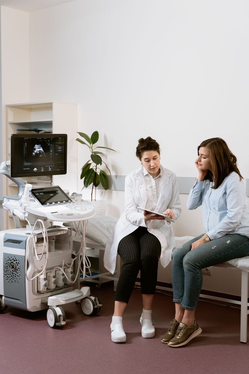
Biochemical prenatal screening.
What prenatal biochemical screening is, when and why it is done, and how to verify the result
Biochemical screening is an analysis of a set of serum markers of pregnancy that makes it possible to estimate the likelihood of a number of conditions developing in the fetus.
The level of certain hormonal markers in a pregnant woman's blood is closely linked to the baby's development. Biochemical screening measures the levels of hormones that change when the fetus has an accompanying chromosomal abnormality. A screening test only indicates an increased risk of Down, Edwards or Patau syndrome — it does not establish a diagnosis! The test is performed in the first trimester of pregnancy.

Prenatal biochemical screening in the 1st trimester is performed at 11-14 weeks of pregnancy. Before the test, an ultrasound is done to determine the gestational age. If the gestational age is determined incorrectly, the test will give an erroneous result. If biochemical screening shows a high probability of chromosomal abnormalities, the doctor may order repeat biochemical screening in the second trimester or invasive diagnostics to verify the diagnosis.
Indications for biochemical screening.
Even young and healthy parents have a risk of having a baby with chromosomal abnormalities, so biochemical screening is recommended for every woman regardless of age and risk group. Groups for whom the test is especially recommended:
- • Mother's age over 35, father's age over 45;
- • Genetic abnormalities in the parents or their blood relatives;
- • A previous child born with birth defects;
- • Consanguineous marriage.
- • Unfavorable history: miscarriages, missed and regressing pregnancies;
- • Infectious diseases contracted before 10-11 weeks of the current pregnancy;
- • Taking medications that are prohibited during pregnancy in the first trimester.
Contraindications to biochemical screening
- • Excess or insufficient body weight of the pregnant woman;
- • Multiple pregnancy;
- • Diabetes mellitus in the mother-to-be;
- • Pregnancy resulting from IVF.
These conditions affect the levels of the hormonal markers being tested, so biochemical screening will give an incorrect result.
There are many software systems for calculating risks.
The main and most widely used of them are the ASTRAIA and PRISCA software systems.
By the way, the ASTRAIA program will most likely be introduced in Kyrgyzstan.
The ASTRAIA program
The ASTRAIA program is the only international program for prenatal diagnosis of fetal chromosomal abnormalities.
This procedure is approved by the Fetal Medicine Foundation and is used in the USA and European countries.
The combined ASTRAIA first-trimester program is performed from 11 weeks 1 day to 13 weeks 6 days.
The program includes:
-
Expert-level ultrasound screening, during which the presence of fetal chromosomal markers is assessed (nuchal translucency thickness, nasal bone, tricuspid regurgitation, ductus venosus, facial angle, Doppler of the uterine vessels).
-
Biochemical screening
This test takes into account two indicators: hCG and PAPP (Pregnancy-Associated Plasma Protein A).
- Medical history and anthropometry
The ASTRAIA program makes it possible to calculate the individual risk of having a baby with genetic defects and to determine your specific risk group. The program includes the latest algorithms for determining the risks of trisomy 13, 18 and 21, which have been approved by The Fetal Medicine Foundation.
Only specialists who have received FMF certification have access to the ASTRAIA program. The doctors of the MEDXPERT diagnostic center in Osh have passed FMF certification and have access to the program.

ASTRAIA makes it possible to determine the risk of late pregnancy complications, such as:
- preeclampsia in the third period of pregnancy;
- fetal growth restriction;
- risk of preterm birth.
All stages of the screening take place on the same day. The blood for biochemical screening is given on an empty stomach — 4 hours after the last meal. Before the test, the pregnant woman is allowed to drink only a couple of sips of still water.
The day before the test, the pregnant woman is advised not to eat sweets, spicy or smoked food, and to avoid fried and fatty foods and citrus fruits.
After the screening results are received, an obstetrician-gynecologist should consult the pregnant woman and give recommendations.
Our ultrasound doctors have received the international FMF certificate from The Fetal Medicine Foundation, the world's leading organization in fetal medicine, which means their professionalism is recognized at the international level.
The PRISCA program
The PRISCA program (Prenatal Risk Calculation) is a program that calculates the risk of congenital chromosomal abnormalities in the fetus based on the pregnant woman's blood. In Europe it is the standard for diagnosing congenital fetal abnormalities (Down, Edwards and Patau syndromes).
Down syndrome is a tripling of chromosome 21; as a result, the full set of chromosomes consists not of 46 chromosomes but of 47. It is believed that the older the woman, the higher the risk. The tripling occurs at the moment of conception. Prenatal testing for Down syndrome is carried out in the 1st trimester (chorionic villus sampling) and in the 2nd trimester (amniocentesis). Both procedures are invasive — that is, they involve inserting special instruments into the woman's body cavities, which may carry high risks of complications for the fetus.
Edwards syndrome is an extra chromosome 18. It is a genetic disease caused by a tripling of the 18th pair of chromosomes.
Patau syndrome is a genetic disease characterized by the presence of an extra chromosome 13.
Neural tube defects are the most common congenital malformation, leading to serious fetal abnormalities.
PRISCA prenatal biochemical screening
- is non-invasive;
- causes no complications;
- detects fetal abnormalities.
How is PRISCA performed?
Risk calculation with the PRISCA program is indicated for all pregnant women regardless of age and the outcome of previous pregnancies. It is carried out in the 1st and 2nd trimesters using the biochemical markers typical of each period. In the 1st trimester these are the free beta subunit of hCG and PAPP-A protein together with ultrasound data. In the 2nd trimester — AFP, free estriol, and ultrasound. A standard blood draw is done for the test.
Important: the blood draw and the ultrasound must be done on the same day!
The main difference between the "ASTRAIA" and "PRISCA" programs is that the first program takes into account the probability of early and late preeclampsia — a serious complication of pregnancy.
Reference values for biochemical screening
Each marker has its own reference values, and deviations from them indicate problems. After all the results are interpreted and assessed together, taking into account the mother-to-be's age, the overall risk of chromosomal abnormalities in the fetus is determined.
How can the diagnosis be confirmed?
The only way to confirm the diagnosis is invasive diagnostics, in which the abdominal wall is punctured and fetal genetic material is collected for testing. In the first trimester, chorionic villus sampling is performed; in the second — amniocentesis and cordocentesis.
Invasive diagnostics gives a 100% reliable result, but carries a number of risks that are very low in percentage terms yet become significant given how many pregnant women are tested.
Increasing the informativeness of first-trimester screening
It is possible to use a more accurate screening test, namely non-invasive prenatal testing.
Non-invasive prenatal test - NIPT
Having a healthy baby is the primary goal of any woman. There are genetic disorders that are incompatible with life and/or cause suffering to both the child and the parents. Detecting a genetic abnormality early in pregnancy is very important. For this purpose, laboratory practice has long used non-invasive biochemical screenings (PRISCA, ASTRAIA), which calculate the risk of a genetic abnormality based on blood test results and ultrasound data. These are affordable biochemical tests, and they are always done for women registered at a women's clinic. The result of such a test is a calculated figure showing the risk of having a baby with a genetic abnormality for a woman of a certain age with such hormone levels and ultrasound data.
If this calculated risk turns out to be high, the woman is referred for an invasive test - amniocentesis.
Biochemical screening programs are not highly accurate; they are based on a combination of statistical data, hormone levels and fetal size on ultrasound.
Amniocentesis is the most accurate method, but it is invasive (the amniotic sac has to be punctured to obtain fetal cells for testing) and carries a risk of complications and pregnancy loss.
Medical science has not stopped searching for new screening tests that would be more accurate and not dependent on calculated indicators. Recently, NIPT (non-invasive prenatal tests) have proven themselves well as screening tests: reliable, convenient and not interfering with the normal course of pregnancy. The accuracy of the method reaches over 90%, since it examines the genetic material of the fetus (its DNA) in the venous blood of the mother-to-be.
How is this possible? Scientists have found that starting from about 10 weeks of pregnancy, fetal DNA circulates freely in the woman's blood. Thanks to modern technologies, doctors have learned to isolate and examine it, detecting the most common chromosomal changes.
Thus, almost every woman can give a venous blood sample once she reaches 10 weeks of pregnancy and find out the genetic health of her future baby.
In which cases is NIPT testing most useful?
• If biochemical screening (PRISCA or ASTRAIA tests) has shown a high risk of chromosomal abnormality
• In pregnant women over 35 years of age. At this age, all methods based on calculated statistical data give a high risk of genetic abnormality, since the program takes into account the statistical age-related risk.
• If genetic disorders were found in the fetus in previous pregnancies
• If the woman wants to be tested specifically by this method. Keep in mind that standard biochemical screening can be done free of charge, covered by compulsory medical insurance (OMS), at a women's clinic. NIPT is a rather expensive test that can only be done for a fee at commercial laboratories.
In which cases is NIPT not possible?
• If the pregnancy is less than 10 weeks
• More than 2 fetuses
• There are signs of a missed singleton pregnancy
• An organ or tissue transplant, including bone marrow, was performed before the pregnancy
• In the presence of cancer
So, non-invasive prenatal tests can be performed in singleton and twin pregnancies. If the pregnancy is a natural singleton pregnancy or occurred after IVF using the woman's own egg, all NIPT tests are available. In other cases there are restrictions.
What are the differences between the tests in the NIPT range?
NIPS T21 - diagnosis of Down syndrome only. The test detects an extra chromosome 21 if the fetus has one. Down syndrome is considered one of the most common chromosomal abnormalities, and its frequency increases with the woman's age. It is performed in pregnancies resulting from natural conception, IVF with the woman's own egg or with a donor egg; in singleton and twin pregnancies, as well as in surrogacy and when one embryo in a twin pregnancy has been reduced.
NIPS 5 – a DNA test for 5 syndromes – a non-invasive test for 5 syndromes that can detect abnormalities of chromosomes 13, 18, 21 and, in most tests, abnormalities of the sex chromosomes X and Y.
• Down syndrome (chromosome 21)
• Edwards and Patau syndromes (extra chromosome 18 and 13, respectively)
• Klinefelter syndrome (extra X chromosome) - possible in boys
• Turner syndrome (missing X chromosome) - occurs only in girls
NIPS 5 is universal: it can be performed both in a natural singleton pregnancy and in a twin pregnancy, when carrying a donor egg, for surrogate mothers, and when one fetus in a twin pregnancy has been reduced.
3 basic test panels:
• NIPT Panorama, basic panel - in addition to a natural singleton pregnancy, it can be performed in a twin pregnancy if both embryos are developing; for women carrying a donor egg and in surrogacy. The test distinguishes the zygosity of twins (monozygotic or dizygotic). The test is considered complete if chromosomes 13, 18, 21 have been examined.
• NIPT Harmony, basic panel – also available in singleton and twin pregnancies, includes determination of twin zygosity, in IVF using a donor egg and in surrogacy.
• NIPT Panorama, basic panel - differs from the other basic panels in that it is used only in a natural singleton pregnancy or IVF with the woman's own egg.
The basic panels make it possible to detect abnormalities of fetal chromosomes 13, 18, 21, X and Y, as well as triploidy.
• Triploidy (an extra set of chromosomes) – leads to severe multiple defects incompatible with life
• Jacobs syndrome (an extra Y chromosome is detected) – only in males, leads to infertility
• XXX syndrome (an extra X chromosome)
• Sensorineural hearing loss – develops with a 50% probability if one of the parents has a dominant gene
NIPS is unique not only in its clinical significance but also in its accessibility. Singleton pregnancy, twin pregnancy (with determination of zygosity), including when one of the embryos in a twin pregnancy has been reduced. In IVF with a donor egg and in surrogacy this test is not appropriate, because mutations associated with genetic diseases must be determined from the blood of the woman whose egg gave rise to the embryo.
The following 2 panels include microdeletion syndromes:
• NIPT Panorama, extended
• NIPT Panorama – DNA test for 18 syndromes
Microdeletions are breaks in a segment of chromosomes; they are less common but no less dangerous, and they cannot be suspected on ultrasound.
• DiGeorge syndrome – congenital immunodeficiency, heart defects and facial deformities.
• 1p36 deletion syndrome – severe intellectual disability due to defects in brain development.
• Cri-du-chat (cat's cry) syndrome – severe intellectual impairment, visual disorders and abnormalities of the larynx.
• Angelman syndrome – known as "happy puppet syndrome", manifests with seizures, chaotic movements, frequent laughter for no reason
• Prader-Willi syndrome – resembles Down syndrome in its features
Can the sex of the fetus be determined, and in which cases?
Yes, at the woman's request any NIPT determines the sex of the fetus, and this is available in both singleton and twin pregnancies.
Please note that results indicating risks of pathological syndromes require a consultation with a geneticist and additional invasive diagnostics.
Invasive diagnostics.
Invasive tests such as chorionic villus sampling and placentocentesis are considered the gold standard for diagnosing fetal chromosomal disorders. Their accuracy is 99%.
In which cases is invasive diagnostics prescribed?
Screening results (ultrasound and biochemical blood test) in the first trimester of pregnancy may show an increased risk of fetal developmental disorders: more than 1%. This by no means means that the baby will be born ill, but it gives the doctor grounds to refer the mother-to-be for additional examination.
A patient may also be referred for invasive diagnostics if the screening showed good results. This means she has other indications for such an examination.
Indications for invasive prenatal diagnostics:
• Children with chromosomal or other birth defects have already been born in the family
• One of the parents has been diagnosed with a balanced chromosomal rearrangement (that is, there is something unusual in the structure of the chromosomes, but it has not led to developmental disorders)
• Ultrasound markers of chromosomal abnormalities were found during screening
• Biochemical screening showed an increased risk of having a baby with a chromosomal disease, for example, Down syndrome.
Why is it important to detect chromosomal diseases as early as possible?
Chromosomal defects cannot be cured. Their effect on fetal development is irreversible. Down syndrome, which everyone knows, is one example. But there are even more severe chromosomal disorders.
Timely diagnosis of fetal malformations makes it possible to learn about the likelihood of having an unhealthy child and helps the future parents understand whether to continue the pregnancy. Invasive diagnostics is carried out as prescribed by a doctor.
How is invasive diagnostics performed?
According to modern standards of prenatal diagnosis, it is carried out using invasive methods, that is, it requires intervention in the woman's body.
Invasive diagnostics is carried out in laboratory conditions. Under ultrasound guidance, the doctor makes a puncture in the anterior abdominal wall and removes a few chorionic villi (or a tiny piece of the placenta, if the placentocentesis method is used).
The obtained material is studied in the laboratory by counting the number of chromosomes in the cells. Since chorionic cells are identical to fetal cells, after the count the doctor can say precisely whether the baby's chromosomes are all right.
Sterile instruments, disposable needles and gloves are used during the tests. Therefore, the risk of introducing an infection is practically excluded.
Likelihood of complications after invasive diagnostics
As with any other invasive procedure, complications can occur.
These include increased uterine tone and preterm birth.
The risk of complications ranges from 0.3 to 1%. It should also be noted that in modern clinics doctors do everything to avoid any possibility of complications. In addition, as a precaution, invasive fetal diagnostics is performed only when indicated.
This information is for informational purposes only.
Do not self-diagnose or self-medicate
consult specialists.
MEDXPERT Diagnostic Center (ultrasound), Osh, 2022