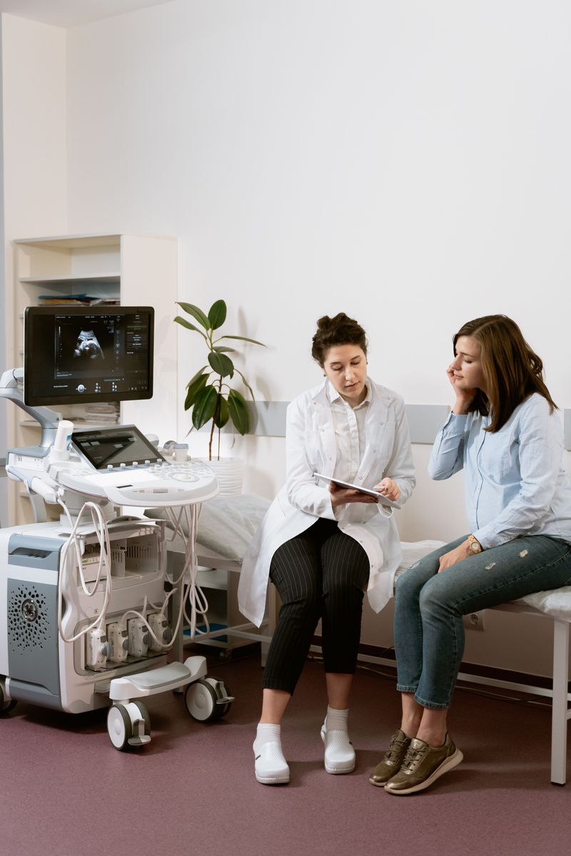
Ultrasound of the lower limb vessels is a method using ultrasound waves that allows the vessels to be displayed graphically and the parameters of their condition to be assessed.
To analyze the characteristics of blood flow, the ability of an ultrasound wave to produce an image when reflected from moving blood cells is used. This technique is called Doppler measurement, or Doppler ultrasonography.
Types of Lower Limb Vascular Ultrasound
- USDG (two-dimensional Doppler ultrasonography)
This examination is now used rather rarely, due to the emergence of other types of leg vessel ultrasound, as well as devices with a higher diagnostic level. This type of examination can be used to assess the patency of veins and the condition of the valves found in the superficial, deep and perforator veins (provided the valves are in their typical location).
- Duplex scanning (duplex angioscanning)
This is a combination of Doppler examination and power Doppler mapping. Put simply, during this examination, areas with different blood flow velocities are highlighted in different colors.
Duplex ultrasound of the lower limb vessels is the "gold standard". Only on the basis of this examination (in addition to their own physical examination) can a doctor make a well-founded diagnosis and prescribe treatment. It makes it possible to assess:
the condition of the walls of the arteries and veins
the patency of both deep and superficial veins
the nature of damage to the vein valves and the degree of their insufficiency, wherever the veins are located
the presence of blood clots, their type and size, and the degree to which they narrow the vessels
and also to determine the cause of recurrence of varicose vein disease after surgery or sclerotherapy.
- Triplex ultrasound of the vessels and veins of the lower limbs
This is a color examination of the vessels – the optimal examination method for patients who have problems with the arteries or veins of the legs. It is especially informative when the plan for upcoming surgery needs to be drawn up as precisely as possible, to avoid recurrence or complications after the operation.
Who Needs This Examination
The arteries of the legs should be examined:
-
if you have diabetes mellitus
-
if you suffer from high blood pressure, whatever its cause
-
if walking is accompanied by leg pain
-
if you have leg pain at night, especially when it eases after lowering your legs off the bed
-
if you are overweight
-
if you smoke
-
if your feet get cold quickly even at a normal ambient temperature
-
if you have a history of myocardial infarction
-
if you have had surgery on the leg vessels
-
if your blood cholesterol level is elevated
Doppler Ultrasonography of the Venous Collectors of the Lower Limbs Is Performed in the Following Cases
-
swelling of the legs, especially appearing towards evening
-
visibly varicose veins
-
leg cramps
-
if pregnancy has been complicated by dilated leg veins, swelling or pain
-
leg pain, especially if accompanied by an increase in local or general body temperature
-
changes in the color of the skin on the legs
-
trophic ulcers.
Preparing for the Procedure
Doppler ultrasonography of the lower limbs is performed without any prior preparation. It requires neither a special diet nor stopping the medications used to treat your arteries or veins. If you wear compression garments, you will need to take them off for the examination.
How a Leg Ultrasound Is Performed
The vessels are first examined with the patient lying down, legs bent at the knees. The doctor must then also examine the vessels with the patient standing upright.
Before starting the examination, a small amount of special gel is applied to the legs; it serves to eliminate interference caused by air getting directly under the probe.
During the procedure, the doctor manually selects the ultrasound frequency depending on the depth of the vessels and the required level of detail. A frequency of 6-12 megahertz is most often used. Deep veins are examined with low-frequency probes.
How USDG Results Are Interpreted
It is best to have a leg vessel ultrasound done not where you are simply handed a report with numbers and a brief conclusion, but where the vessels are examined directly by a phlebologist or vascular surgeon. Based on the data obtained, they will assess the characteristics of blood movement through the vessels and then describe the further treatment approach.
The following parameters are used to assess blood flow in the arteries:
Vmax – the maximum blood flow velocity in the artery, recorded during systole
Vmin – the minimum blood flow velocity, recorded during diastole
RI – vascular resistance: the ratio of the difference between the systolic and minimum blood flow velocities to the maximum velocity
PI – pulsatility index, determined almost like RI, but a more sensitive indicator of changes in the vessel lumen
thickness of the inner and middle layers of the vessel wall (IMT).
ABI: the ratio of the systolic pressure in the tibial artery to the same pressure in the brachial artery. It should be approximately 1.0 or slightly higher.
There are no such numerical parameters for veins. During USDG of the limb vessels, the price of which varies between clinics, the doctor assesses the patency of the venous collectors of the lower leg — deep and superficial — as well as the largest veins running through the abdominal and pelvic cavities — the iliac veins and the inferior vena cava.
All possible blood clots are visualized, and the valve system and the condition of the communicating veins are assessed. Tests are also performed with raising the leg and compressing the veins with a tourniquet, which gives an idea of the direction and nature of blood flow and the presence or absence of pathological reflux of blood in the opposite direction.