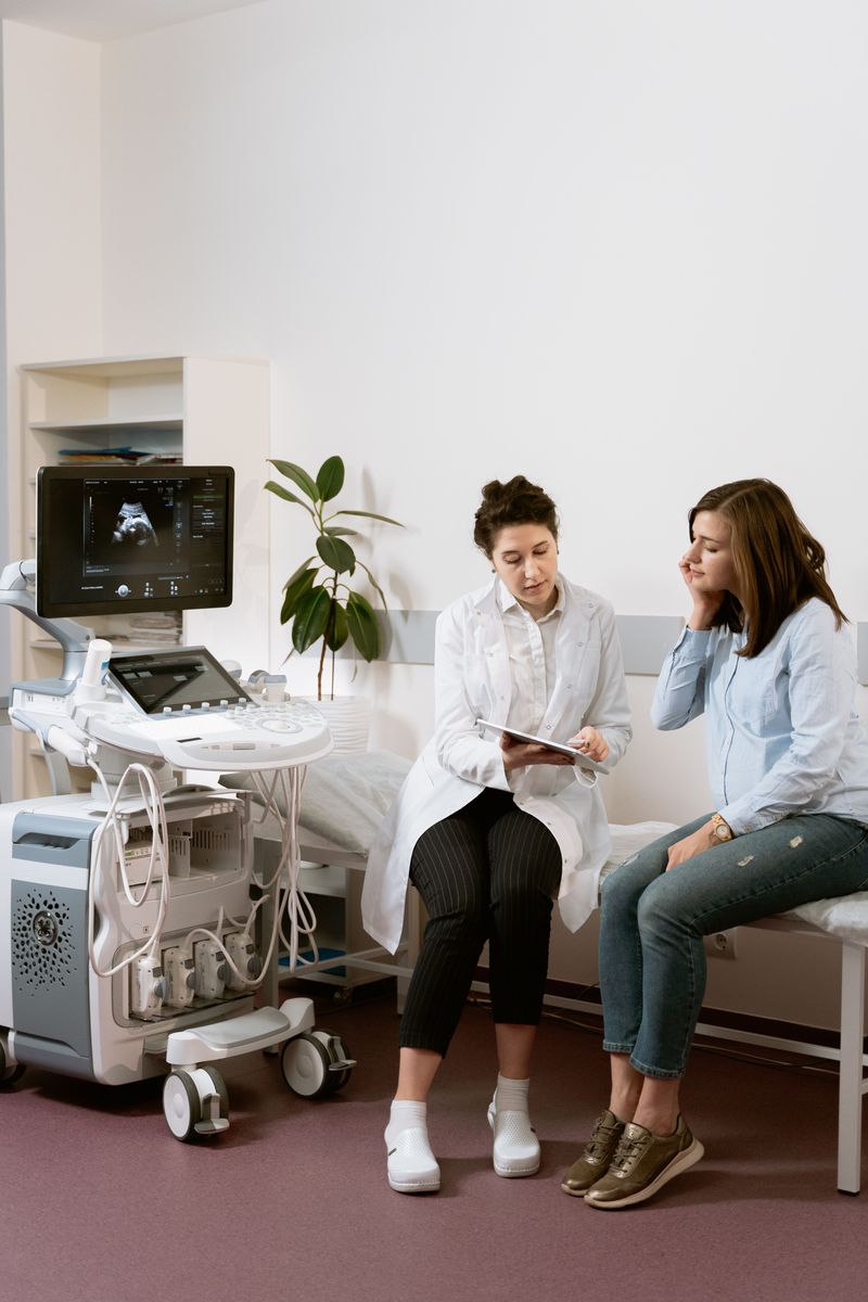
Duplex scanning of blood vessels is one of the methods for examining veins and arteries based on the use of ultrasound. It makes it possible to track changes in the condition of the vessel walls and to observe blood flow at any point in a vessel.
The design of the examination device itself is based on the principle of echolocation. The working surface is designed so that it emits and captures ultrasound pulses. In fact, this is how a two-dimensional image of the vessel is created from the data obtained.
Today, duplex scanning of blood vessels is one of the most accurate and safest methods for examining them. The use of ultrasound is completely harmless; in addition, the device can be set up to examine a wide variety of vessels, both superficial and the deepest ones (the aorta, coronary arteries, kidney vessels).
Duplex scanning: how the procedure is performed. The whole process is completely safe and painless and does not require any special preparation. First, the patient lies down on a couch in a comfortable position, with the head slightly raised. The doctor performing the scan applies gel to the device's probe so that it can move easily and freely over the patient's skin. The information received by the probe is converted into a digital signal and then displayed on the monitor screen. The procedure itself usually lasts about half an hour, but keep in mind that much depends on the size of the area being scanned.
Indications for the procedure. It cannot be said that this examination method is suitable for everyone. In some cases, an ordinary blood pressure measurement or a cardiogram is enough. However, in the presence of serious diseases, ultrasound duplex scanning will provide the most complete information about the condition of blood vessels and veins.
There are a number of symptoms which, when detected, are a reason to order an ultrasound of the blood vessels:
- Headache;
- Dizziness;
- Ringing in the ears;
- Memory decline;
- Unexplained restlessness and anxiety.
All of this indicates impaired blood circulation in the vessels of the neck and head. If such symptoms are detected, duplex scanning of the head vessels should be done without delay, because lack of treatment can lead to complications.
The symptoms listed above can accompany a whole range of diseases. For example, if a patient has poor vessel patency (in hypertension), vasospasm occurs, that is, the vessels narrow. In a disease such as atherosclerosis, plaques form on the vessel walls.
Duplex scanning of blood vessels. In cervical osteochondrosis, due to displacement of the vertebrae and bone overgrowths, the vertebral artery may be compressed. In this case, duplex scanning of the neck will be highly relevant. This is far from a complete list of diseases that can have similar symptoms.
Duplex scanning of the neck and head vessels is an accessible and, most importantly, safe ultrasound method that helps diagnose what caused a particular disease.
The vessels involved in supplying blood to the brain should be examined especially carefully. Transcranial duplex scanning, an ultrasound examination of the intracranial vessels, helps with this.
Impaired blood supply can lead to various diseases, and therefore the need for duplex scanning of the carotid arteries, which do not always receive proper attention, becomes especially pressing.
Duplex scanning of blood vessels makes it possible to detect changes in hemodynamics, to identify organic and functional disorders of blood flow, as well as preclinical signs of disease.
Duplex scanning of veins and arteries
It is worth saying that vein diseases have become widespread nowadays. Duplex scanning of veins, the price of which depends on many factors, is in greater demand than ever.
The methods of modern phlebology today provide an excellent opportunity to stop the disease and eliminate cosmetic defects. However, is it worth letting the illness reach a critical state if there is a chance to diagnose it at its very earliest stage? The sooner a patient undergoes ultrasound duplex scanning of the veins, the sooner he can start treatment, and therefore get rid of these unpleasant problems.
The most common vein diseases include varicose disease, venous angiodysplasias and post-thrombotic disease. The first step towards getting rid of a disease of this kind is a consultation with a specialist and duplex scanning of the veins.
In some cases, a specific type of examination is needed. For example, if a patient is troubled by throbbing pain in the temple area, frequent dizziness, darkening of vision, as well as "floaters" or "ripples" before the eyes, then duplex scanning of the brachiocephalic arteries should be performed.
In addition, if a patient experiences a brief loss of consciousness or any other neurological symptoms, the best option in this situation will be duplex scanning of the head. This examination should also be done if the patient has been diagnosed with vegetative-vascular dystonia or chronic fatigue.
With frequent complaints of pain in the calf muscles, cramps, numbness in the legs, trophic changes in the skin of the legs, changes in skin color on the lower limbs, or swelling syndrome, duplex scanning of the vessels of the lower limbs should be done. It can be used to diagnose varicose disease and thrombosis of the superficial and deep veins of the lower limbs. Duplex scanning of the arteries of the lower limbs makes it possible to diagnose aneurysms, arteriovenous fistulas, and abnormalities in the structure and course of the arteries.
It also becomes possible to study the elasticity and thickness of the vessels and to visualize the valve apparatus. At the end of the examination, the doctor will have complete information about the condition of the vessels, after which timely treatment is prescribed.
Duplex scanning of arteries and veins is one of the popular methods of Doppler ultrasound, which makes it possible to obtain information about the state of blood flow in the major arteries and veins.
Duplex and triplex scanning
Based on duplex scanning, another diagnostic method was created — triplex examination. It is the same duplex scanning, but supplemented with color Doppler mapping. That is, the capabilities of ordinary duplex scanning are expanded by encoding information using color.
Duplex and triplex scanning performs functions such as examining the anatomy of the vessels and assessing vessel patency and blood flow. Thanks to this, the doctor can obtain more accurate information about the condition of the vessels.
Most often, color duplex scanning is ordered to examine the arteries of the head, neck, limbs and abdominal organs.