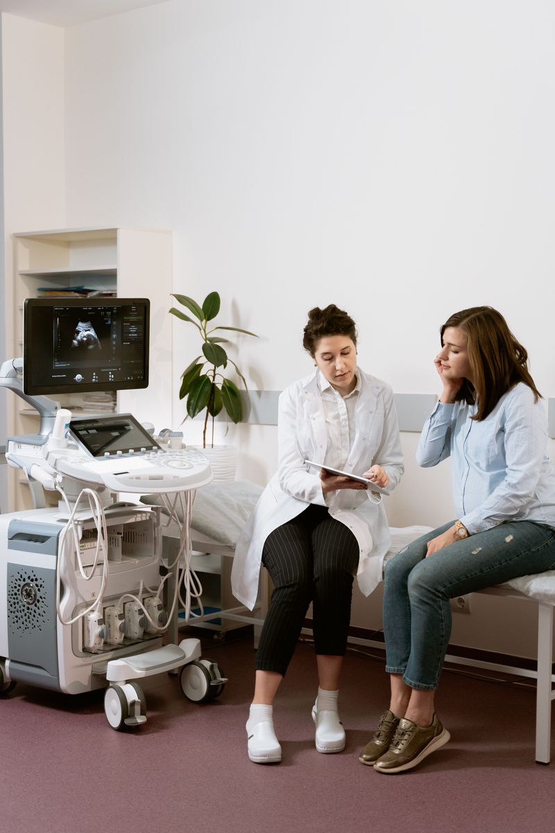
Description of the method. Who needs an abdominal ultrasound and why.
This is a very informative and safe examination method that allows the condition of the internal organs to be determined with high accuracy – to assess the size and structure of the organs examined, the features of their shape and position, and developmental anomalies, and to detect areas of inflammation, degenerative changes in the tissues of internal organs, vascular disorders, benign or malignant tumors, and stones in the gallbladder and bile ducts. It is perhaps the most accessible and highly informative diagnostic method in gastroenterology. It is especially important for detecting gallstone disease, inflammatory diseases of the gastrointestinal tract (cholecystitis, pancreatitis, etc.), and liver cirrhosis.
Ultrasound is safe, so it can be repeated several times at short intervals. All organs are examined "through" the abdominal wall, and the examination takes 15-20 minutes on average.
With this ultrasound diagnostic method, even the smallest areas of change can be found, and many diseases can be detected at an early stage, when there are no clinical symptoms yet.
Despite the high informative value of abdominal ultrasound, the possibility of false-positive and false-negative results should be taken into account. That is why it is essential to have laboratory tests done and to interpret the ultrasound results together with the findings of other diagnostic methods and the clinical picture of the disease.
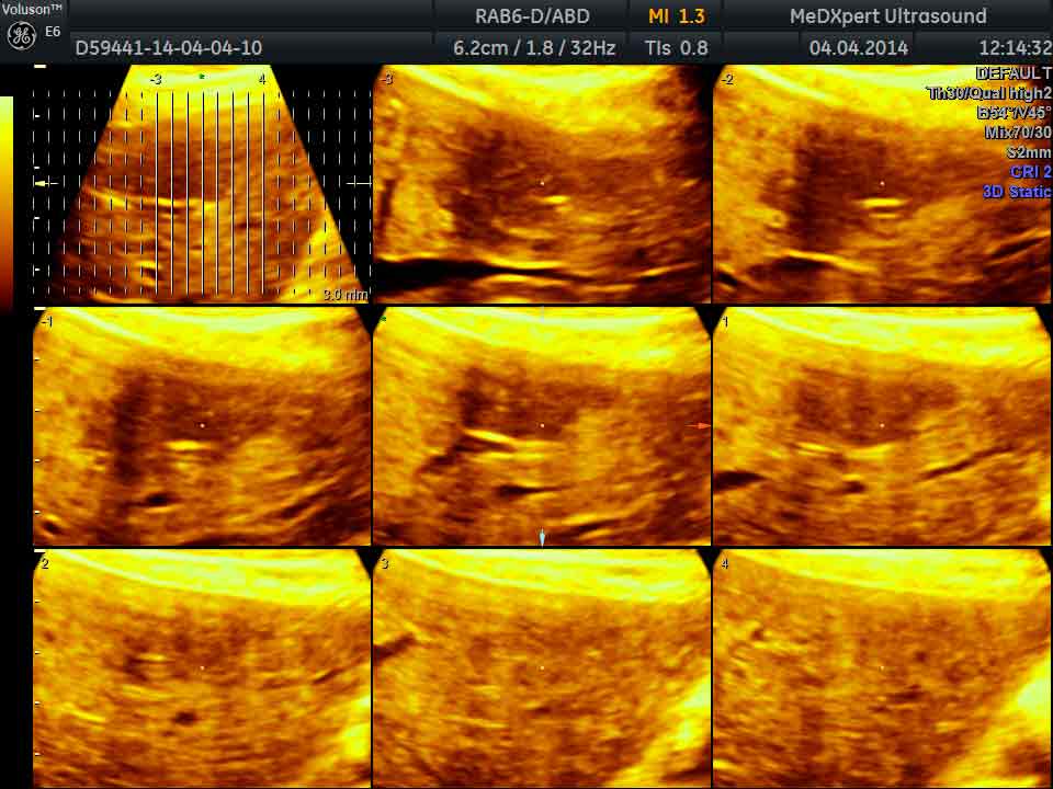
Liver ultrasound is a diagnostic method that reveals changes developing in the liver tissue as a result of various diseases (liver enlargement, changes in the structure of the liver tissue, growth of connective tissue, appearance of tumors, etc.).
Gallbladder ultrasound shows its position, size, shape, outer and inner contours, the thickness and structure of its walls, the presence of additional inclusions in its cavity, and its emptying function. It helps detect shape anomalies, gallbladder dyskinesia, acute and chronic cholecystitis, stone formation in the gallbladder and its ducts, polyps and other lesions.
Pancreatic ultrasound involves assessing the size and shape of the organ in different views and evaluating its condition and the changes in its structure caused by an ongoing disease process. It helps diagnose acute and chronic pancreatitis, detect pancreatic cysts and tumors, and diabetes mellitus.
Spleen ultrasound is usually ordered when developmental defects are suspected (absence of the organ, abnormal position, altered shape), in blood diseases, or in spleen injury, which is common in abdominal trauma.
When should an abdominal ultrasound be done?
- abdominal pain in any location;
- increased gas (bloating);
- a bitter taste in the mouth; a feeling of heaviness under the right ribs;
- digestive problems: belching, heartburn, constipation, diarrhea, nausea, reduced appetite, weight loss or weight gain;
- problems with the skin of the face or body (various rashes, redness, telangiectasias, itching);
- suspected benign or malignant tumors;
- chronic diseases of the digestive organs;
- after abdominal injuries;
- when preparing for surgery, and after surgery on the abdominal organs
Abdominal ultrasound shows:
The size, shape and structure of the liver, cirrhosis, enlargement, fatty degeneration of the liver (fatty liver disease).
Stones in the gallbladder, inflammation of the gallbladder itself (cholecystitis), or blockage of the bile ducts.
Changes in the size of the spleen (both enlargement and reduction) and any lesions in it.
Diseases of the pancreas (diffuse enlargement due to swelling during inflammation, diffuse or focal changes in its structure).
Tumors of the abdominal organs.
Bruising of internal organs after an injury.
Free fluid in the abdominal cavity (ascites).
An abdominal aortic aneurysm, which may appear as a pulsating tumor-like mass in the abdomen (this would be a reason for further examination).