Description of the method. When it should be done.
You can book a breast ultrasound at the MEDXPERT Diagnostic Center (Ultrasound) in Osh. This will help you and your attending doctor save time in making a diagnosis and starting treatment.
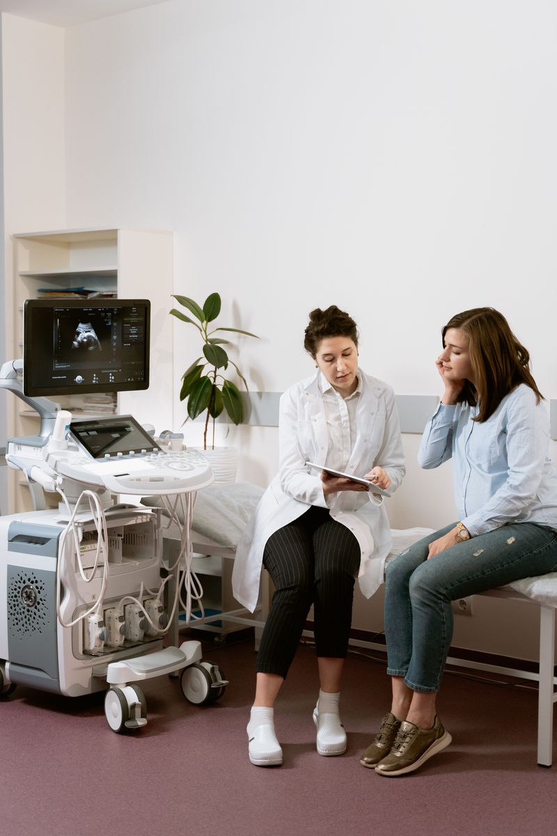
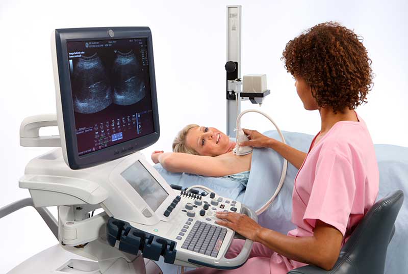
Ultrasound at MEDXPERT is performed on high-end and expert-class General Electric Voluson scanners, with high-frequency, high-resolution probes. The devices are equipped with elastography technology (measuring the elasticity and stiffness of tissue), which makes it possible to predict with high probability whether a lesion in the breast is malignant.
Breast ultrasound is prescribed to detect abnormalities and as a preventive measure, so as not to miss the onset of a disease. Painless and harmless, the method makes it possible to detect possible changes at early stages.
Indications for breast ultrasound:
- signs of inflammation (pain, swelling or redness) of the breast
- discharge of various kinds from the breasts
- breast injuries and chest bruises
- a suspected lump found on palpation or as a result of mammography
- to monitor and check the size of existing cysts
- postpartum mastitis
- accompanying gynecological and endocrine problems
- to check atypical mammography results
- follow-up examinations for women on long-term hormonal treatment
- hormonal contraception
- for women who have reached the age of 30, as a preventive examination
- regular follow-up after breast surgery
- if you have silicone breast implants (to assess their condition)
- if you have mastopathy, for monitoring every six months
In men, breast ultrasound makes it possible to answer the question of what is causing breast enlargement — excess fatty tissue or the presence of true female glandular tissue (true gynecomastia).
Breast ultrasound is an effective and accurate method for diagnosing diffuse breast diseases.
Ultrasound is the main method for diagnosing breast diseases in women under 40. For women over 40, X-ray mammography is recommended, due to differences in breast structure at different ages.
During breast ultrasound, the condition of the axillary (armpit) lymph nodes is assessed at the same time, which helps the mammologist decide on treatment.
Preparing for a Breast Ultrasound
No special preparation is required, but it is best done in the appropriate phase of the menstrual cycle. The optimal time for a breast ultrasound is the first half of the cycle (up to day 12). In the second phase of the cycle, the breasts are more sensitive, as they undergo hormonal changes that can make the examination more difficult. If your cycle is irregular or you do not have periods, the examination can be done on any convenient day.
If a lesion in the breast is suspected, ultrasound is performed regardless of the day of the menstrual cycle.
Mammography or Breast Ultrasound — Which Is Better?
Advantages of ultrasound:
- a non-invasive, painless procedure without X-ray radiation;
- it can produce a clear image of structures that cannot be seen on mammography;
- it is performed in real time;
- it helps in the differential diagnosis of various pathological lesions;
- it can be used in different age groups;
- it can be used to examine pregnant and breastfeeding women;
- it is the method of choice for examining breasts with diffuse mastopathy;
- the gland can be examined in multiple positions.
Disadvantages of ultrasound:
a repeat examination or a biopsy (aspiration or needle) may be required.
Advantages of mammography:
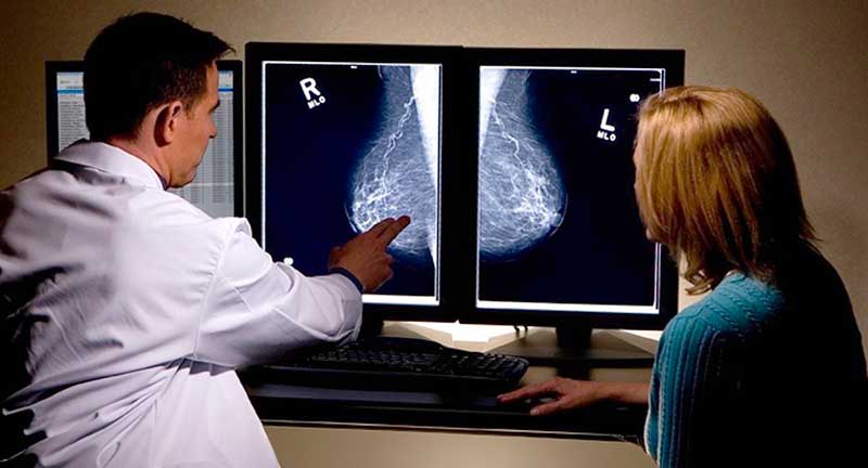
- the ability to detect relatively early forms of breast cancer, including minimal and non-palpable lesions 1–3 mm in diameter; the ability to assess how far the process has spread and the growth pattern of the tumor;
- the ability to examine the gland in different projections;
- the ability to perform a full differential diagnosis of nodular lesions and intraductal diseases;
- having the images makes it possible to monitor the condition of the breasts over time and, if necessary, adjust treatment in a timely manner.
Disadvantages of mammography:
- X-ray radiation is used, which makes it impossible to perform the examination on pregnant and breastfeeding women;
- an ultrasound may be needed to check its atypical results;
- for some women the examination is painful;
- the examination is less informative in the presence of diffuse mastopathy.
Important!
Different methods of breast examination can be used together, complementing each other. Nor should you neglect self-examination, which doctors recommend doing monthly (after the end of each period). Self-examination should begin by checking the symmetry of the breasts, the condition of the nipple (whether it is retracted, whether there is any discharge) and the quality of the skin; then each breast is palpated clockwise, starting from the upper outer quadrant. If you have any doubts — be sure to see a doctor.
How Often Can a Breast Ultrasound Be Done?
Ultrasound is a harmless, painless, highly informative examination method that has no contraindications. Breast ultrasound can be prescribed to pregnant women and children, as ultrasound does not damage body tissues.
The examination takes 15 minutes. The ultrasound doctor's report is issued immediately after the examination is completed.